
TOP STORIES • JUN 29 2012
TRENDING TOPICS: CYBERPUNK • GALAXY S III • WINDOWS PHONE 8


4:18PM on Dec 20, 2010


11 cheap gifts guaranteed to impress science geeks

Science comes up with a lot of awesome stuff, and you don't need a Ph.D, a secret lab, or government funding to get your hands on some of the coolest discoveries. We've got a list of 11 mostly affordable gifts that are guaranteed to blow your mind, whether or not you're a science geek.
Click on any image to see it enlarged.

Aerogel isn't just neat, it's useful. It supports up to 4,000 times its own weight and can apparently withstand a direct blast from two pounds of dynamite. It's also the best insulator in existence, which is why we don't have Aerogel jackets: it works so well that people were complaining about overheating on Mt. Everest.
Price: $35

EcoSpheres came out of research looking at ways to develop self-contained ecosystems for long duration space travel. They're like little microcosms for the entire world, man. But ask yourself: are we the shrimp, or the algae?
Price: $80

Every once in a while, a meteorite smashes into Mars hard enough to eject some rocks out into orbit around the sun. And every once in a while, one of these rocks lands on Earth. It doesn't happen often, but it does happen, and whoever finds the meteorite is allowed to cut it up into bits and sell it to people who want to have their very own piece of another planet.
Price: $70+

The existence of a shape with these properties was conjectured in 1995, but it took ten years for someone to figure out how to actually make one that worked. And then everyone was embarrassed when it turned out that turtles had evolved this same basic shape in their shells a long time ago, to make it easier for them to roll themselves back over if they get flipped.
Price: $150

So what's next year's new color going to be? It's looking like orange, but they're not quite what I'd call affordable yet. Something to look forward to for next year, especially if you're going for your own personal laser rainbow.
Price: $110

While you probably shouldn't lick your fingers after playing with it, gallium isn't toxic and won't make you crazy like mercury does. And if you get tired of it, you can melt it onto glass and make yourself a mirror.
Price: $80

The upshot of this effect is that some things you eat taste spectacularly different. Straight Tabasco sauce tastes like donut glaze. Guinness tastes like a chocolate malt. Goat cheese tastes like cheesecake. After about an hour of craziness, your taste buds go back to normal, no harm done.
Price: $15

While the technology hasn't reached the point where you can affordably get a complete sequence of an entire genome, looking at specific markers is still good enough to suggest some things worth looking out for while spurring a lively nature versus nurture debate.
Price: $100

The difference between the models and the real thing is that by adding an extra dimension, you can make it so that the neck of the bottle doesn't actually intersect the side of the bottle. Take a couple aspirin and try to picture that in your head.
Price: $35

These giant plushes are the perfect way to make the holidays even more awkward, when you present your friends with a variety of adorable STDs. Microbiologists, at least, will appreciate that they're more or less anatomically correct, too.
Price: $9

Ferrofluids are found in everything from speakers to hard drives, but it's much more fun to play with when when you've got a puddle of it naked and out in the open.
Price: $40
Like this list? Check out our Holiday Gift Guide, which is an easy place to find all of our holiday gift lists for 2011.

1. Aerogel
Also known as frozen smoke, Aerogel is the world's lowest density solid, clocking in at 96% air. It's basically just a gel made from silicon, except all the liquid has been taken out and replaced with gas instead. If you hold a small piece in your hand, it's practically impossible to either see or feel, but if you poke it, it's like styrofoam.Aerogel isn't just neat, it's useful. It supports up to 4,000 times its own weight and can apparently withstand a direct blast from two pounds of dynamite. It's also the best insulator in existence, which is why we don't have Aerogel jackets: it works so well that people were complaining about overheating on Mt. Everest.
Price: $35
2. EcoSphere
Inside these sealed glass balls live shrimp, algae, and bacteria, all swimming around in filtered seawater. Put it somewhere with some light, and this little ecosystem will chug along happily for years, no feeding or cleaning necessary, totally oblivious to the fact that the rest of the world exists outside.EcoSpheres came out of research looking at ways to develop self-contained ecosystems for long duration space travel. They're like little microcosms for the entire world, man. But ask yourself: are we the shrimp, or the algae?
Price: $80
3. Mars Rock
NASA has been trying to figure out how to get a sample of rock back from Mars for a while now. You can beat them to the punch and pick up a little piece of the red planet without having to travel a hundred million miles, by just taking advantage of all the rocks Mars sends our way.Every once in a while, a meteorite smashes into Mars hard enough to eject some rocks out into orbit around the sun. And every once in a while, one of these rocks lands on Earth. It doesn't happen often, but it does happen, and whoever finds the meteorite is allowed to cut it up into bits and sell it to people who want to have their very own piece of another planet.
Price: $70+
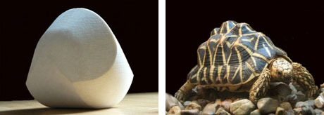
4. Gömböc
The Gömböc is a self-righting object, which means that no matter which way you put it down, it stands itself back up. It's like a Weeble, except it doesn't cheat by having a weight at the bottom, and it's the only shape that can do this.The existence of a shape with these properties was conjectured in 1995, but it took ten years for someone to figure out how to actually make one that worked. And then everyone was embarrassed when it turned out that turtles had evolved this same basic shape in their shells a long time ago, to make it easier for them to roll themselves back over if they get flipped.
Price: $150
5. Violet Laser Pointer
It's no longer geeky enough to have a red laser pointer, or a green laser pointer, or even a blue laser pointer. Keep moving up the spectrum until you get to violet, and you'll find the new hotness at 405 nanometers.So what's next year's new color going to be? It's looking like orange, but they're not quite what I'd call affordable yet. Something to look forward to for next year, especially if you're going for your own personal laser rainbow.
Price: $110
6. Gallium
Gallium is a silvery metal with atomic number 31. It's used in semiconductors and LEDs, but the cool thing about it is its melting point, which is only about 85 degrees Fahrenheit. If you hold a solid gallium crystal in your hand, your body heat will cause it to slowly melt into a silvery metallic puddle. Pour it into a dish, and it freezes back into a solid.While you probably shouldn't lick your fingers after playing with it, gallium isn't toxic and won't make you crazy like mercury does. And if you get tired of it, you can melt it onto glass and make yourself a mirror.
Price: $80
7. Miracle Berries
By themselves, Miracle berries don't taste like much. The reason to eat them is that they contain a chemical called miraculin that binds to the sweet taste receptors on your tongue, changing their shape and making them respond to sour and acidic foods.The upshot of this effect is that some things you eat taste spectacularly different. Straight Tabasco sauce tastes like donut glaze. Guinness tastes like a chocolate malt. Goat cheese tastes like cheesecake. After about an hour of craziness, your taste buds go back to normal, no harm done.
Price: $15
8. DNA Genotyping
There's nothing more personal than someone's own DNA. And there are ways to give the gift DNA that won't get you children or arrested. With just a little bit of spit, you can get an genotype analysis that will reveal fun insights about longevity, intelligence, susceptibility to diseases, and even food preferences.While the technology hasn't reached the point where you can affordably get a complete sequence of an entire genome, looking at specific markers is still good enough to suggest some things worth looking out for while spurring a lively nature versus nurture debate.
Price: $100
9. Klein Bottle
If you want to give a mathematician something to try to wrap their head around, a Klein bottle is a good place to start. A real Klein bottle is an object with no inside and no outside that can only exist in four dimensions. These glass models exist in three, which means that unlike the real thing, they can actually hold liquid.The difference between the models and the real thing is that by adding an extra dimension, you can make it so that the neck of the bottle doesn't actually intersect the side of the bottle. Take a couple aspirin and try to picture that in your head.
Price: $35
10. Giant Plush Microbes
They're cute! They're fuzzy! They're potentially deadly! All of the microbes, bacteria, and viruses that you know and love (or maybe not) are available in huggable forms about a million times larger than real life. In the picture are gonorrhea, syphilis, mono, and herpes.These giant plushes are the perfect way to make the holidays even more awkward, when you present your friends with a variety of adorable STDs. Microbiologists, at least, will appreciate that they're more or less anatomically correct, too.
Price: $9
11. Ferrofluid
Magnetic particles suspended in oil never looked so sexy. That's all a ferrofluid is, and it looks pretty gross until you put it in close proximity to a magnet, at which point it grows spikes all over the place as the fluid flows out along magnetic force lines.Ferrofluids are found in everything from speakers to hard drives, but it's much more fun to play with when when you've got a puddle of it naked and out in the open.
Price: $40
Like this list? Check out our Holiday Gift Guide, which is an easy place to find all of our holiday gift lists for 2011.
For the latest tech stories, follow us on Twitter at @dvice
DVICE CATEGORIES
3D Tech Android Announcements Apparel Apple Apps Art & Design Buildings Camcorders Cameras Car Electronics Cellphones CES 2010 Cloud Computing Computer Peripherals Downloading DVICE Gift GuideDVICE TV DVICE Video E-Readers Editorial Feature Feature Future Tech Futurism Galleries Gaming Gift Guide Google GPS Green Tech Green Week HDTV Home Theater Household Infographics InternetiPhone Kitchen Lists Medical Microsoft Military Miscellaneous Office Outdoor PCs Polls Portable Gadgets Quizzes Reviews Robots Science Security Software Space Tablets Tech In-Depth Top Stories Toys Vehicles Video Wi-Fi

MOST COMMENTED
3 drawbacks that could spoil Microsoft's Surface tablet at launch
How to reverse Faceook's decision to force its email on you
By 2050, washing machines will levitate

Editor:
Kevin Hall
editor(at)dvice.com
Contributing Editors:
Evan Ackerman, Features
Raymond Wong, Reviews
Evan Dashevsky
Eileen Marable
Michael Trei
Megan Wollerton
Stewart Wolpin
International Editor:
Adario Strange
Kevin Hall
editor(at)dvice.com
Contributing Editors:
Evan Ackerman, Features
Raymond Wong, Reviews
Evan Dashevsky
Eileen Marable
Michael Trei
Megan Wollerton
Stewart Wolpin
International Editor:
Adario Strange


